


The disease is poorly understood, although several thousand observations with subsequent diagnoses and treatments have been explained.
The many variations and uncertainties of the clinical picture of varicose veins in the small pelvis cause major errors in the diagnosis, which in the future affect the consequences.
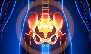
Characteristics of small pelvic varicose veins
Pelvic veins are several times longer than arteries, which results in greater capacity. This is due to the phylogenesis of the vascular system of the pelvic area. Pelvic veins are very adaptable and potentially prone to remodeling, which contributes to the formation of interconnected tissues.
The speed and direction of blood flow are regulated by valves, which are controlled by complex humoral mechanisms. The valve balances the pressure in different parts of the venous network.
When the valve stops performing its function, blood stagnation develops, this causes vascular pathology and the formation of varicose veins. The uniqueness of the pelvic vein lies in the fact that the broad uterine ligament, which maintains a wide vessel lumen, can narrow it, causing pathology.
Cause
Pathological pelvic vein dilation may be due to the following reasons:
- Disorders of blood outlets;
- Removal of venous stems;
- Compression of the collateral trunk by changing uterine position, for example, in retroflection;
- ovarian vein valve deficiency (congenital or acquired);
- Obstructive postphlebitic syndrome;
- Connective tissue pathology;
- Arteriovenous angiodysplasia;
- Sitting long, hard physical work;
- Varicose veins in the lower leg;
- Pregnancy (3 or more) and childbirth (2 or more);
- Diseases of the female genital area (chronic salpingo-oophoritis, ovarian tumors, uterine fibroids and genital endometriosis);
- Pelvic organ attachment;
- Obesity.
Classification by disease level
By enlarging the veins, the following degrees are distinguished:
- to 0, 5 cm, "corkscrew" vessels;
- 0, 6-1 cm;
- more than 1 cm.
Diseases of the disease
- varicose veins perineum and vaginal anterior space;
- small pelvic vein congestion syndrome;
Symptoms
- The most common - frequent pain in the lower abdomen, perineum after prolonged static and dynamic pressure. Pain increases in the second phase of the cycle, after hypothermia, fatigue, stress, exacerbation of various diseases.
- Feeling "out of place", pain during and after sex.
- Menstruation - menstrual irregularities, including pain.
- Secretions, more than usual, of the genital tract.
- Blood stagnation leads to infertility, miscarriage, abortion.
- Urinary incontinence due to bladder vein expansion.
Diagnostics
Diagnosis of disease only with successful complaints in only 10% of cases.
Palpation of the inner wall of the pelvis, making it possible to feel tight and long vein knots. When viewed in a mirror, cyanosis of the vaginal mucosa can be seen.
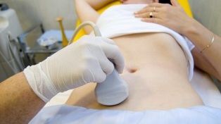
The optional procedure is ultrasound examination with color Doppler mapping, which makes it possible to detect not only varicose veins, but also venous thrombosis, post-thrombophlebitic occlusion. Ultrasound shows turtles, "worm-like", structures without signal reflection, localized on the lateral surface of the uterus.
Doppler effect is based on the blue and red color of venous and arterial blood flow.
A device for ultrasound examination with the help of a special program recognizes the movement of blood from sensors and in other directions, calculating blood flow velocity and channel type.
But the exact definition of a vein or artery remains with the doctor. The Doppler method works in almost all cases, the exception to the rule is determined by our body, because the blood flowing from the heart is not always arteries and vice versa.
Therefore, ultrasound diagnostic doctors look at these arterial or venous channels, their size, the rate of blood flow in them and many indications that are not needed by the average person, but play an important role in making a diagnosis. For this, transabdominal and transvaginal sensors are used.
In 5. 7% of cases, the disease is diagnosed by chance during examination. Usually the diameter of the venous vein is 0. 4 cm.
CT and MRI are very accurate. With this method, it is possible to detect the accumulation of varicose veins in the ligaments of the uterus, ovaries and around these organs. It is possible to determine the corresponding pathology.
A very reliable method is phlebographic research.
Contrast is performed at the height of the Valsalva test, against blood flow. This allows you to see valve failure accurately.
Left rethngenorenoscopy, renal phlebography, superselective phleboovarioscopy and phleboovariography are also used. This method makes it possible to determine hemodynamic and anatomical changes in the kidney veins and the places where the gonad veins flow into them.
Superselective fleboovarioscopy is performed by catheterization of the gonad vein through the femoral or subclavian contralateral vein, followed by contrast injection.
A large amount of blood from the varicose veins of the uvine plexus is excreted through the ovarian veins. But in the case of hypertension, it occurs through the extraorganic uterine veins into the internal iliac veins. Venous plexus, where outflow may occur, including sacral plexus and bladder.
In left-sided phleboovaricography, there are 3 stages of venous stasis in the left ovarian uviform plexus:
- There is no outflow from the left ovarian plexus, or it follows an additional short path.
- There is an extra long road.
- Two additional outflow paths are visible, or one additional and additional route.
In stages 2 and 3, varicose veins of the right ovarian plexus plexus form.
Laparoscopy is used for differential diagnosis. Pathological winding veins are located in the ovarian area, towards the round and broad ligaments. They look like large cyanotic conglomerates with thin, taut walls.
The complexity of the diagnosis lies in the fact that the disease is often hidden behind the signs of inflammatory processes, differs in clinical manifestations, disguises as endometriosis, prolapse of internal organs, postoperative neuropathy and many extragenital diseases.
Treatment
The main goal of treatment is to remove reflux in the veins. In the early stages of the disease, conservative treatment is used. In the final stages of the disease, surgery is the treatment of choice.
Conservative treatment

This consists in normalizing venous tone, improving hemodynamic and trophic processes.
Symptomatic treatment for individual symptoms. Non-steroidal anti-inflammatory for pain, for bleeding - hemostatic therapy.
The main drugs in conservative treatment are venotonic drugs and antiplatelet agents.
Phlebotonics - improves vascular wall tone and improves blood flow. With this disease, it is better to consult a gynecologist about certain medications.
Physiotherapy is an important method.
Surgical Treatment
- Varicose vein resection.
- Shunting gonado-caval.
- Sclerosis in laparoscopy.
- ovarian vein occlusion using X-ray endovascular method.
People's solution
Since the main factor in the onset of the disease is the weakness of the valve apparatus, all folk remedies used for varicose veins in the lower part of the leg are also used for this pathology.
The most commonly used are: common hazel, hop, nettle, horse chestnut, dandelion root, kombucha, willow, oak, St. John's wort. John's wort, string, pollen and many more plants.
Effective are: treatment with bath with oak, chestnut, willow, chamomile, pharmacy, dried herbs, St. John's wort. John's wort, string.
Prevention
- The first thing to do if you have a complaint, prognosis or illness listed above is to contact your gynecologist.
- It is necessary to normalize the work regime and rest, trying not to be in an upright position for long periods of time, physical stress.
- Perform exercises to prevent "pedals", "stand-birch", "scissor leg"
- Follow a diet: eat foods high in vitamins E, P, C, try to eat only white meat, less fatty meats, replace with fruits, vegetables, cereals.
- Drink plenty of fluids, but not less than 1. 5 liters a day.
- Lose weight, bad habits.
- Talk to your doctor about wearing compression clothing, this will increase blood flow from the bottom of the leg, thus reducing congestion in the pelvis.
- Avoid baths, saunas, steam rooms, hot showers.
In order not to get sick with a disease that is difficult to diagnose, you need to follow the precautionary recommendations listed above. Pamper your health as the most precious thing in life.
For suspicious symptoms that you can not get rid of in a few days, you must see a doctor. He must give you qualified help and save you from suffering.